Akrotome Imaging | Moving the Future Closer
Advances in cancer diagnostics have enabled the detection of cancer at its earliest stages when the chances for successful intervention are highest. For many tumors, surgery offers the best option for a cure, with the potential to remove all cancer tissue from the patient (a “complete resection”).
However, the full potential of surgery is not realized because, in many cases, it is difficult for surgeons to see where cancer tissue ends and healthy tissue begins. Akrotome (a combination of two Greek words that means “cutting at the edge”) is focused on the development of fluorescent molecular probe technologies that “light up” cancer, enabling clear and precise visualization of tumors, with emphasis on tumor margins, where the cancer is growing rapidly and where invasion into healthy tissue occurs.
Akrotome provides surgeons with the visualization tools they need to guide them to achieve complete resection of cancer tissue.
Who We Are
Akrotome is a Delaware corporation, first incorporated late 2008; the current team has been in place since 2012.
Our Focus
We are dedicated to the clinical translation of cancer-targeted molecular probes into enhanced approaches for surgery and diagnostics, leading to improved outcomes and patient quality of life while positively impacting healthcare economics.
How We Are Different
Akrotome’s molecular probes were designed from the ground up with an emphasis on the real-world requirements of cancer surgeons and the clinical workflow in the Operating Room. The result is a family of probes that offer superior performance, economics, and flexibility to other molecular probes on market.
While other probes use a “one size fits all” approach that requires systemic administration of large doses of probe hours or even days before surgery, Akrotome’s probes can meet a surgeon’s preference for an individual surgery, supporting the following application methods:
- In vivo with topical administration
- In vivo with systemic administration
- Ex vivo with topical administration
In addition, Akrotome probes are quenched and do not activate until they encounter cancer, virtually eliminating the background fluorescence common with other probes.
This flexibility translates into real and quantifiable benefits, including: reduced regulatory risk and expense; faster time to market, and potential lower-per-use cost/enhanced profitability
Akrotome Officers
 Brian Straight, PhD., CEO
Brian has 20 years experience in image processing technologies & research/clinical informatics. He has founded and led a number of successful companies in the clinical imaging and informatics space. Brian also has deep experience in validated Q&A, strategic marketing, sales, and operations management.
 Matthew Bogyo, PhD., Co-Founder
Matt is a Stanford University Professor in the Departments of Pathology and Microbiology & Immunology. He received his Ph.D. from MIT in 1997 and became a Full Professor at Stanford in 2013. One of his major accomplishments has been to develop fluorescently quenched probes that activate upon binding to an active protease. The cysteine cathepsins targeted by these probes are proteases that have been shown to have roles in various aspects of immune cell function and are also important regulators of multiple disease pathologies. His work has demonstrated the utility of Activity Based Probes for imaging tumor margins, tumor response to chemotherapy, atherosclerosis, asthma and pulmonary fibrosis.
 James Basilion, PhD., Co-Founder
Jim is a Professor at Case Western Reserve University in the Departments of Radiology and Biomedical Engineering. After a stint in the private sector, Jim joined the faculty at Harvard Medical School as an Assistant Professor of Radiology. He was then recruited by the Case Western Reserve University as an Associate Professor and Director of the National Foundation for Cancer Research (NFCR) Center for Molecular Imaging As a tenured Full Professor in the Departments of Radiology and Biomedical engineering, Jim is working to develop a vital clinical translational infrastructure to service the Center’s molecular imaging pipeline.
Scientific Advisory Board
 Sunil Singhal, MD.
Dr. Singhal is Director of the Thoracic Surgery Research Laboratory and is William Maul Measey Associate Professor in Surgical Research at Penn Medicine (the University of Pennsylvania Health System). He received his MD from the University of Pennsylvania and completed his residency at the Johns Hopkins Hospital in Baltimore. Dr. Singhal’s practice is focused on lung cancer and esophageal cancer, including complex resections and advanced disease. He performs minimally invasive (VATS and robotic) pulmonary surgery. Dr. Singhal is the Director of the Thoracic Surgery Research Laboratory and his interest lie in development of advanced technology for intraoperative imaging to detect small quantities of disease and immunotherapy to treat patients with advanced lung and thymic cancer. He is conducting several clinical trials to improve care for patients with advanced disease.
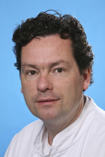
Alexander L. Vahrmeijer, MD, Ph.D.
Dr. Vahrmeijer is a surgical oncologist and one of the world’s leading experts on the clinical translation of fluorescence imaged-guided surgery technologies. He received his MD and Ph.D. in Leiden and specialized in colorectal cancer surgery and the treatment of liver metastases. He now heads the Image-Guided Surgery Group at Leiden University Medical Center (LUMC). In 2015, he was awarded the prestigious Bas Mulder Award of the Dutch Cancer Society. His current research focus is on the clinical introduction of novel probe technologies like Akrotome’s FIRE probes.


