
Akrotome Molecular Probes Offer a Precisely-Targeted Intervention for Cancer Surgery
Achieving a surgical cure for cancer depends on “getting it all” – removing all diseased tissue from a patient. To meet this need, Akrotome has developed a family of quenched, substrate-based probes (qSBPs) that precisely target enzymes produced by malignant tumors.
What is a qSBP?
A qSBP is a molecular probe that targets a specific molecule—in this case enzymes produced by many cancers called cathepsins. You can think of the probe like a flashlight hidden under a dark cloth. When the probe reaches the tumor, it binds to any active cathepsins it finds. Upon binding, the “cloth” is pulled from the flashlight and the probe glows brightly, enabling the surgeon to see the tumor and its margins clearly and making it easier to see all the tissue that needs to be removed.
How are Akotome’s Probes Different?
Other probes require large IV doses of the probe to be given to the patient by IV hours or even days before a procedure. Akrotome’s probes do not require systemic administration, but can be used in the OR by applying the probe topically when needed either to the surgical cavity or directly to tissue that has been removed from the patient. Akotome’s probes activate in minutes, rather than hours, and precisely define both the tumor and its margins.
Akrotome FIRE Advantages
- Rapid Activation
- Exceptional signal-to-background ratio
- Flexible Administration
– IV Systemic
– Topical in vivo
– Topical ex vivo - Integrates seamlessly into the OR workflow
Akrotome Mission Statement
Akrotome Imaging Inc. is driven to revolutionize the management of diseases by development of innovative molecular imaging technologies.
Superior Probes. Unmatched Flexibility
Unlike competitive probes that require the administration of significant doses hours before surgery and may lack precision in visualizing diseased tissues, Akrotome’s probes have a flexibility that is unmatched. While the probes can be administered by IV, they may also be used topically either for the inspection of the margins on cancer tissue that has been removed from a patient (ex vivo), or to evaluate a tumor and its margins within the patient during surgery (in vivo).
Only Akrotome’s smart probes activate rapidly, diffuse well through tissue, irrespective of vascularization. They provide an unmatched capability for precise, global evaluation of the full surface area of both the tumor and tumor cavity, enabling rapid, intraoperative assessment of margin status, dramatically increasing the potential for a surgical cure.
Defining The Cutting Edge
One of the biggest challenges cancer surgeons face is the detection of disease in the marginal tissues surrounding a tumor. This tissue may contain tissue from the main tumor, as well as tiny cancer “seeds” that may be difficult or impossible to detect using vision or touch. Complete removal of a tumor requires that the surgeon resects not only the main tumor itself, but also any disease in the tissue margins around the tumor. A complete resection means that these marginal tissues are free of disease (a clean margin).
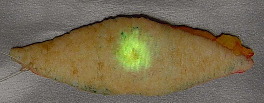
Sample from an ongoing clinical trial demonstrating the power of FIRE to discriminate between cancer and surround normal tissues for non-melanoma skin cancer resection.
Akrotome’s probes are small molecules that target enzymes produced by cancer called cathepsins. While cathepsins are produced in the main bulk of the tumor itself, they are even more prevalent at the tumor margins. The probes are like tiny flashlights covered in a dark cloth. When they encounter the cathepsins, they bind to the enzyme, the “dark cloth” is removed, and the probe glows brightly in a near-infrared wavelength of light.
Depending on how the probe is used, the surgeon can either examine a tumor after it is removed to determine if there is cancer on or near its surface, or he or she can look into the patient using a small, near-infrared camera to detect and remove any glowing tissue.
Real-Time Precision
Studies indicate that outcomes for cancer patients who are candidates for surgery are closely correlated with the completeness of surgical resection. For most surgeries, the determination of whether all cancer tissues have been removed is made by pathological examination after the surgery is over. In cases where pathology finds cancer in marginal tissues, patients may require more aggressive postoperative treatment. In some cases, patients may even be recalled for additional surgical procedures.
Our probes are highly specific. They target tumor tissue and activate quickly with a bright signal, enabling a surgeon to see the tumor and its margins with precision while the surgery is taking place. Such guided surgery has the potential to result in more complete surgical resections, with the potential for positive impacts for patients and for the economics of healthcare.